This research:
- Revealed associations between nicotine, alcohol, and marijuana use during two periods of adolescence and smaller gray matter volume in two brain areas at age 25.
- Supports a hypothesis that as the brain matures during adolescence, it passes through stages in which it is particularly sensitive to substance exposure.
Dr. Michael Windle at Emory University and colleagues from other institutions performed magnetic resonance imaging of the brains of 110 25-year-old African Americans. The young men and women had participated in a study of Strong African American Families, a program designed to reduce the impact of childhood poverty on rural African Americans. During the study, they had reported their substance use to researchers annually from age 11 to 21.
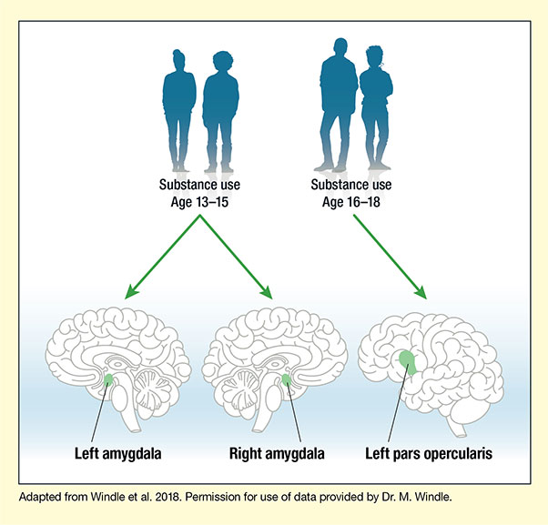
The researchers found that higher levels of alcohol, cigarette, and marijuana use before age 19 correlated with smaller gray matter volume in two brain areas (see Figure). The amygdala was smaller in youths who had reported higher use of the substances at ages 12 to 15. The pars opercularis, a subregion of the inferior frontal gyrus, was smaller in those who reported higher use of the substances at ages 16 to 18. To the researchers’ knowledge, theirs is the first study to show a relationship between substance exposure and the pars opercularis.
The location and timing of the observed associations parallel the staged maturation of the adolescent brain, in which emotional circuits come fully online prior to cognitive circuits. The amygdala is part of the limbic system, which matures earlier and is strongly implicated in emotional responses, especially fear. The pars opercularis is part of a forebrain circuit that matures later and supports the ability to refrain from impulsive behaviors.
Less gray matter in an area may indicate that it contains fewer neurons, which suggests a reduced capacity to shape behavior. Dr. Windle and colleagues say that relatively low gray matter volume in the two areas may be a contributing cause or an effect of substance use, depending on whether it:
- Was already present before the adolescents used the substances, in which case it may have rendered them more likely to become users or continue using after an initial exposure, or
- Came about because the drugs inhibited the developmental expansion of gray matter volume in the affected areas.
Both etiologies might also pertain. To resolve the issue, researchers will need to conduct a longitudinal study in which they obtain brain images before adolescence, collect substance use data throughout adolescence, and obtain follow-up images after adolescence.
The researchers also looked at gray matter volume in the ventral striatum. They found no link between substance exposure and gray matter volume in that area, but speculate that a link might emerge in a study with more participants.
Dr. Windle says, “When confirmed and extended, the findings may facilitate an understanding of brain regulatory processes underlying risky behaviors, including substance use, that may serve to guide targeted preventive interventions.”
This study was supported by NIH grant DA027827.
- Text Description of Figure
-
The figure illustrates the relationship between substance use during adolescence and reduced volumes of certain brain areas during young adulthood (age 25). At the top, dark blue silhouettes represent the age groups when substance use was assessed. The two figures on the left represent participants who used substances during early adolescence (age 13–15), and the two figures on the right represent participants who used substances during middle adolescence (age 16–18). At the bottom are three drawings of the brain to indicate the locations of the brain regions analyzed. The left and middle drawings show lengthwise cross-sections of the brain, with the front of the brain facing left in the left drawing and facing right in the middle drawing. Green ovals deep inside the brain and just above the brainstem illustrate the location of the left amygdala in the left illustration and of the right amygdala in the middle drawing. The drawing on the right shows an outside view of the brain from the side, with the front of the brain facing left. A green shape slightly forward and below the center of the brain indicates the location of the left pars opercularis. Two green arrows pointing from the left set of silhouettes to the left and middle brain drawings indicate that substance use at age 13–15 was associated with significantly reduced volume of the left and right amygdala. A green arrow pointing from the right set of silhouettes to the right brain drawing indicates that substance use at age 16–18 was associated with significantly reduced volume of the left pars opercularis.
Source:
- Windle, M., Gray, J.C., Lei, K.M., et al. Age sensitive associations of adolescent substance use with amygdalar, ventral striatum, and frontal volumes in young adulthood. Drug and Alcohol Dependence 186:94-101, 2018.