
This study found that:
- Mice genetically engineered to lack mu opioid receptors in the medial habenula were less sensitive to the unpleasant effects of the opioid antagonist naloxone and showed fewer naloxone-induced withdrawal symptoms.
- The modified mice still experienced the normal locomotor, pain-relieving, and rewarding effects of morphine.
Mu opioid receptors (MORs) are known to mediate opioid drugs’ pleasant and reinforcing effects. A NIDA-sponsored study by Dr. Laura-Joy Boulos from the McGill University Douglas Research Centre and her colleagues recently found that MORs located on certain cells in the brain’s “aversion center,” the medial habenula, may also regulate unpleasant, or aversive, effects. The researchers showed that these MORs were responsible for behaviors and withdrawal symptoms induced by the opioid antagonist naloxone.
“This is a very new role for the MOR, classically known as a reward-producing receptor through disinhibition of dopamine neurons at the level of mesolimbic networks,” explains the study’s senior investigator, Dr. Brigitte Kieffer. “Our data show that this receptor would also limit aversion at the level of another brain circuit involving the habenula, suggesting that the MOR is not only ‘pro-reward,’ but also ‘anti-aversion’,” she adds.
The medial habenula is a brain region centrally involved in processing unpleasant experiences. Yet this region also has the highest density of MORs in the brain, which generally process reward-related signals. To investigate whether MORs are also involved in aversion processing, the research team used a targeted genetic manipulation in mice to delete MORs from a subset of neurons that are mainly found in the medial habenula. MOR expression in all other brain regions remained intact. Subsequently, both the modified mice and mice serving as controls that expressed MORs normally throughout the brain were treated with morphine and/or its antagonist, naloxone, and their responses to both drugs were analyzed.
Morphine had the same behavioral and pain-relieving effects in the modified mice as in the control group, and all animals experienced morphine’s rewarding effects (see Figure 1). The responses to naloxone, however, differed between the two groups. In contrast to the control group, the mice lacking MORs in the medial habenula no longer avoided the place where they had received naloxone (a measure of naloxone’s aversive effect). Additionally, morphine-dependent modified mice exhibited fewer physical signs of naloxone-induced withdrawal (see Figure 2). Explains Dr. Kieffer, “This means that a large part of naloxone’s aversive effect is taking place at the level of MORs in the habenula in the brain.”
But how exactly do MORs in the medial habenula contribute to the interplay of rewarding and aversive effects? Various brain regions assign value to environmental stimuli by determining whether the stimuli are rewarding and should be approached or are aversive and should be avoided. Among these, the medial habenula encodes aversive experiences. Researchers also know that, as a general rule, MOR activation inhibits neuronal activity. Dr. Kieffer and her team therefore hypothesized that MORs in the habenula normally inhibit the activity of habenular neurons, keeping this brain structure as inactive as possible. This would limit aversion or aversive states (see Figure 3).
Although clear differences exist between the brains of mice and humans, the human habenula is known to influence drug addiction. Therefore, the results of this study may have implications for aversion elicited by naloxone in humans as well.
This study was supported by NIDA grant DA005010.
- Caption and text description of Figure 1
-
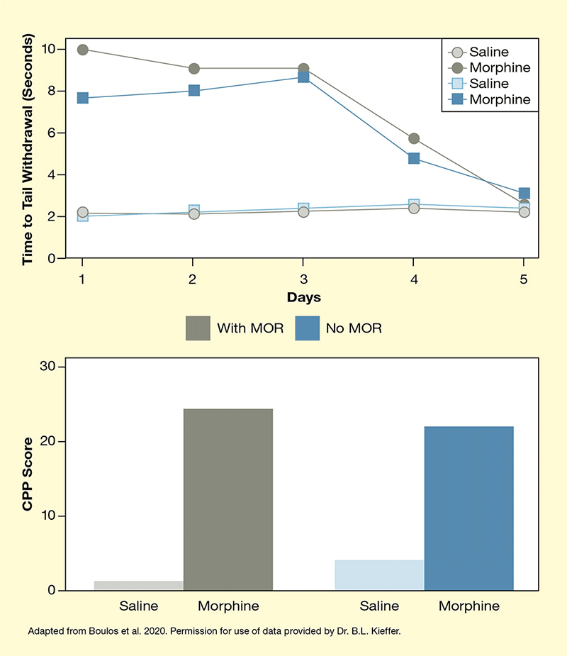
Caption - (Top) To measure the pain-relieving effects of morphine, the researchers measured how long the animals would leave their tail in warm water before withdrawing it with and without morphine treatment. (Bottom) To determine morphine’s rewarding effects, they calculated a conditioned place preference (CPP) score that reflects how much more of their time the animals spent in an area where they had previously received morphine. For both measures, no significant differences existed between mice with and without habenular MORs
Text Description - The two graphs illustrate the role of MORs in the medial habenula in mediating the pain-relieving and rewarding effects of morphine. Gray curves and bars indicate control mice with intact MORs; blue curves indicate mice with no MORs in the habenula. (Top) The line graph shows the pain-relieving effects of morphine and saline in mice with and without MORs in the habenula over time. The animals were treated with saline (open symbols) or morphine (closed symbols) The horizontal x-axis shows days of treatment from 1 to 5; the vertical y-axis shows pain relief expressed as the time after which the mice withdraw their tail from warm water from 0 to 10 seconds. For saline-treated mice with or without MORs, time to tail withdrawal on all days was about 2 seconds. Morphine-treated control mice withdrew their tail after about 10 seconds on Day 1, about 9 seconds on Days 2 and 3, about 5.5 seconds on Day 4, and about 3 seconds on Day 5. Similarly, morphine-treated mice without MORs withdrew their tail after about 7.5 seconds on Day 1, about 8 seconds on Day 2, about 2.5 seconds on Day 3, about 4.5 seconds on Day 4, and about 3 seconds on Day 5. (Bottom) Morphine’s rewarding effects were determined using a conditioned place preference (CPP) score reflecting how much more of their time the animals spent in an area where they had previously received morphine. The vertical y-axis shows the CPP score on a scale from 0 to 30. For saline-treated control mice (left bar), the CPP score was about 1, for morphine-treated control mice (second bar from the left) it was about 24, for saline-treated mice lacking the MOR (second bar from the right) it was about 4, and for morphine-treated mice lacking the MOR (right bar) it was about 22.
- Caption and text description of Figure 2
-
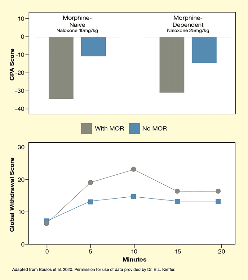
Caption - (Top) To determine naloxone’s aversive effects, the researchers calculated a conditioned place aversion (CPA) score that reflects how much less of their time morphine-naïve and morphine-dependent animals spent in an area where they had previously received naloxone. For both naïve and morphine-dependent mice, the animals lacking MORs showed significantly reduced aversion compared with control animals. (Bottom) Severity of naloxone-induced withdrawal was determined as a global score that included several withdrawal symptoms. Animals lacking habenular MORs had a significantly lower global lower withdrawal score (i.e., exhibited fewer withdrawal symptoms) than did control animals.
Text Description - The two graphs illustrate the role of MORs in the medial habenula in mediating the aversion to naloxone and naloxone-induced withdrawal effects. Gray curves and bars indicate control mice with intact MORs, blue curves indicate mice with no MORs in the habenula. (Top) The bar chart shows the relationship between MOR status and naloxone’s aversive effects, which were determined using a conditioned place aversion (CPA) score reflecting how much less of their time the animals spent in an area where they had previously received naloxone. The vertical y-axis shows the CPA score on a scale from +10 to -40. For morphine-naive control mice (left bar), the CPA score after treatment with 10 mg/kg naloxone was about -35 and for morphine-naïve mice without MORs in the habenula (second bar from the left) it was about -10. For morphine-dependent control mice (second bar from the right), the CPA score after treatment with 25 mg/kg naloxone was about -30, and for morphine-dependent mice without MORs in the habenula (right bar) it was about -15. (Bottom) The line graph shows the relationship between MOR status and naloxone-induced withdrawal. The horizontal x-axis shows time after naloxone administration in minutes from 0 to 20; the vertical y-axis shows severity of withdrawal expressed as a global withdrawal score on a scale of 0 to 30. For the control mice, the global withdrawal score was about 6 at 0 minutes, about 19 at 5 minutes, about 22 at 10 minutes, about 16 at 15 minutes, and about 16 at 20 minutes. For mice without MORs in the habenula, the global withdrawal score was about 7 at 0 minutes, about 13 at 5 minutes, about 15 at 10 minutes, and about 13 at 15 and 20 minutes.
- Text description of Figure 3
-
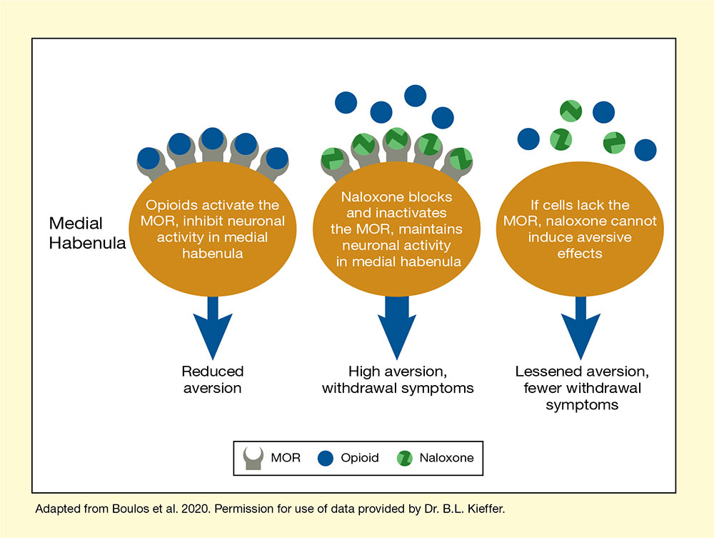
The figure shows a proposed model of how MORs in the medial habenula regulate the aversive and withdrawal-inducing effects of naloxone. Large orange ovals represent the medial habenula. Gray shapes with a circular cutout represent the MORs, blue circles represent opioids, and green circles represent naloxone. Blue arrows pointing downward illustrate the effects of events in the medial habenula on aversion and withdrawal symptoms, with arrow width reflecting aversion/withdrawal symptom severity. The left panel illustrates the normal situation, with opioids binding to and activating the MORs. This inhibits neuronal activity in the medial habenula, resulting in reduced aversion. The middle panel illustrates the effect of naloxone. Naloxone binds to the MOR, displacing the opioids. This inactivates the MOR and maintains neuronal activity in the medial habenula, resulting in high aversion and withdrawal symptoms. The right panel illustrates the effects when cells lack the MORs. Naloxone and opioids cannot bind, and naloxone cannot induce its aversive effects. This results in lessened aversion and fewer withdrawal symptoms.
Source:
- Boulos, L.J., Ben Hamida, S., Bailly, J., et al. Mu opioid receptors in the medial habenula contribute to naloxone aversion. Neuropsychopharmacology. 45(2):247–255, 2020.