
Video length: 1:50
Transcript
[Music]
Dr. Thomas Ross Speaking:
I'm Thomas Ross I'm a staff scientist in the Neuroimaging Research Branch at the National Institute on Drug Abuse.
So our branch uses brain imaging techniques to investigate issues in drug abuse.
Principally we use a technique called functional magnetic resonance imaging. We do it in a way that sensitive to changes in blood flow.
So you can imagine that if part of your brain is working harder then it will need more oxygen so there will be a local increase in blood flow.
We can setup our MRI machine so it is sensitive to those local changes in blood flow.
It shows us what parts in the brain are needed to do certain things maybe if I ask you to make a risky decision I might wanna know what part your bring changes during that risky decision maybe that differs between a drug user and a non-drug user.
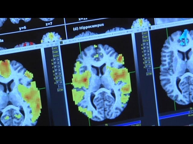
And we will create an image where the color of the image is related to how big the blood flow changes.
Cause we take these images across time hundreds and hundreds and hundreds of images and combine them to make a single image that represents changes related to whatever we're looking at.
When you see these images of colorful, what we call a cluster or blobs region of activation overlaid ontop of a brain those colors represent how big the signal change was.
We hope to better inform the scientific community what parts of the brain are important in critical in drug abuse, that they can be targets for either behavioral therapy or some sort of pharmical therapy to help end drug abuse.